


Dunedin Brain Imaging Study
Overview
To date, brain imaging studies have been limited by the use of small samples that are not representative of the general population, as well as by challenges in successfully connecting brain imaging measures with real-world experiences and outcomes. The Dunedin Brain Imaging Study represents our effort to address these challenges by collecting structural and functional brain imaging measures in the 1000 most studied people in the world. Originally a study of child health, the Dunedin Study has become best known for studying connections between childhood experience and adult health. Now with the help of funding by the US National Institute of Aging, it is becoming a study of aging.
Members of the Dunedin Study include 1,037 people (91% of eligible births; 52% male) born between April 1972 and March 1973 in Dunedin, New Zealand (NZ). The cohort represents the full range of socioeconomic status (SES) in the general population of NZ’s South Island and matches the NZ National Health and Nutrition Survey on key adult health indicators (e.g., body mass index, smoking, GP visits) and the NZ Census of citizens of the same age on educational attainment. The cohort is primarily white (93%), which matches the demographics of the South Island. Assessments were carried out at birth and ages 3, 5, 7, 9, 11, 13, 15, 18, 21, 26, 32, 38, and most recently (completed April 2019) 45 years, when 94% (N = 938) of the 997 Study members still alive took part. Of these Study members, 875 (93% of age-45 participants), who were representative of the original cohort (see figure), completed brain imaging, which started in August 2016 and finished in April 2019.
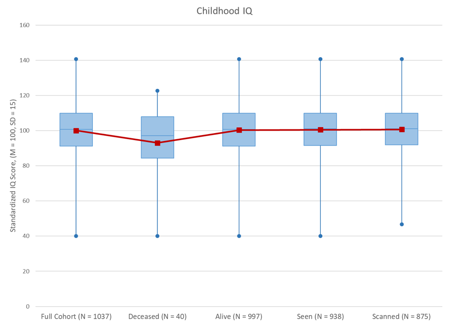
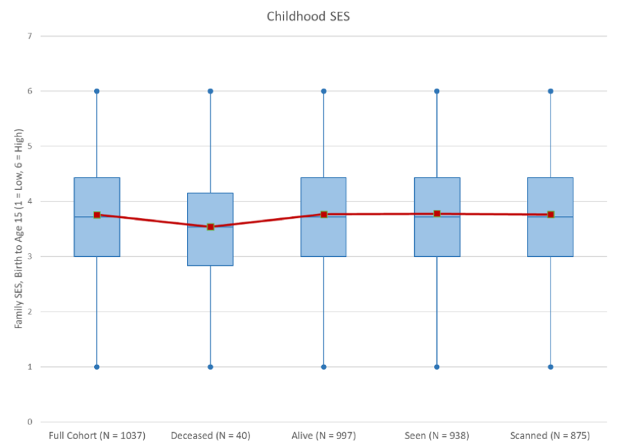 Attrition Analysis. No significant differences in childhood IQ or SES were found between the full cohort, those still alive, those seen at Phase 45 or those scanned at Phase 45. Those who were deceased by the Phase 45 data collection had significantly lower childhood IQ’s than those who were still alive (t = 2.09, p = 0.04), but there were no differences in SES.
Brain Imaging Protocol
The 875 Study members were all scanned using a state-of-the-art Siemens Skyra 3T scanner equipped with a 64-channel head/neck coil at the new Pacific Radiology imaging center in Dunedin.
Structural imaging included a high-resolution T1-weighted MP-RAGE scan (0.9×0.9×1.2 mm), diffusion-weighted scan in 64 directions at 2.5mm isotropic resolution, 3D FLAIR scan (0.9×0.9×1.2 mm), and SWI scan (0.6x0.6x1.6 mm).
Functional imaging was conducted using 2mm isotropic BOLD scans (TR=2s) and included a resting-state (8min 16sec), a face-matching emotion task targeting the amygdala, a color Stroop executive control task targeting the dorsolateral prefrontal cortex, a monetary incentive delay reward task targeting the ventral striatum, and a face-name encoding task targeting the hippocampus.
Brain Imaging Measures
Primary measures generated from the imaging data include global and regional cortical thickness, cortical surface area, grey matter volume, white matter hyperintensities, fractional anisotropy, brain age, and general functional connectivity (based on combined resting-state and task data).
Key People
Ahmad Hariri, Principal Investigator
Terrie Moffitt, Principal Investigator
Avshalom Caspi, Investigator
Richie Poulton, Dunedin Research Unit Director
Annchen Knodt, Research Project Manager
David Ireland, Dunedin Imaging Team Lead
Sandhya Ramrakha, Dunedin Research Unit Manager
Advisory Board
Allen Song, Duke
Wayne Hall, Queensland
Susan Bookheimer, UCLA
Randy Buckner, Harvard
Ranga Krishnan, Rush
Daniel Weinberger, LIBD
Funding
National Institute of Aging: “Neural signatures of healthy and unhealthy aging”
Data Sharing
To inquire about data sharing, email Dr. Hariri (ahmad@haririlab.com).
Publications
For more information on the Dunedin study, visit the Dunedin study website (including a page dedicated to the MRI study) or the Moffitt & Caspi website.